More than 90% of cancer deaths come from metastasis. A new study found that the act of spreading trains cancer cells to spread better, and identified a druggable target at the center of the process

More than 90 percent of cancer deaths are not caused by the original tumor. They are caused by metastasis, by the process through which cancer cells leave their origin, travel through the bloodstream, and establish new colonies in distant organs. A breast cancer patient does not typically die from the mass in her breast. She dies when cancer reaches her liver, her lungs, her brain. Understanding how that journey happens, and specifically how cancer cells survive and improve during transit, is one of the most important unsolved problems in oncology.
A new study from the Chinese Academy of Sciences has identified a mechanism at the heart of that journey that nobody had previously seen: when cancer cells are physically squeezed through the narrow capillaries and tight tissue spaces that they must navigate to spread, the compression itself activates a metabolic program that makes the cells structurally stronger and better equipped to squeeze through the next tight space. The tumor is not simply finding cells that happen to be capable of spreading. The act of spreading is selecting and training them in real time, using the physics of confinement as the trigger.
The researchers identified a single enzyme sitting at the center of this process, showed that blocking it dramatically reduced metastasis in animal models, and confirmed that the same enzyme is elevated in human cancer patients whose tumors have spread. The findings were published in Cell Research.
The bottleneck that kills most cancer patients
To understand why this discovery matters, it helps to understand what cancer cells actually have to do to metastasize. A cell that breaks free from a primary tumor does not simply float through open space to a distant organ. It must squeeze through the dense mesh of extracellular matrix that surrounds tumors. It must force itself into and through blood vessels, which are narrow enough that the cell must deform to fit. It must survive the mechanical stress of being compressed and stretched while circulating. And then it must squeeze back out through capillary walls at a distant site to seed a new tumor.
Each of these steps involves confined migration: the cell moving through spaces smaller than itself, experiencing physical compression that would kill most normal cells. The physical demands of this process are enormous. And yet metastatic cancer cells routinely accomplish it, often becoming more dangerous in the process.
Previous research had established that cancer cells can adapt to mechanical confinement, becoming more capable of navigating tight spaces with experience. What was not known was the molecular mechanism that translates the physical experience of being squeezed into a biological change that makes the cell better at spreading.
The CRISPR screen that found the answer
To identify which metabolic enzymes are specifically required for cancer cells to migrate through confined spaces, the research team led by Weiwei Yang conducted a genome-scale CRISPR screen targeting 1,685 metabolic enzymes simultaneously. They engineered colorectal cancer cells to migrate through microfluidic channels designed to mimic the narrow capillaries that cancer cells traverse during metastasis, then used CRISPR gene deletion to systematically eliminate each of the 1,685 candidate enzymes one at a time, asking which deletions specifically impaired confined migration without affecting normal unconfined movement.
One enzyme emerged from the screen with a clear and specific signal: dihydrolipoamide dehydrogenase, known as DLD, a mitochondrial enzyme involved in energy metabolism that had not previously been linked to cancer spreading.
When the researchers deleted DLD from cancer cells, those cells moved normally in open spaces but became significantly impaired when trying to navigate confined channels. When they restored DLD, confined migration recovered. The enzyme was specifically required for the kind of movement that cancer uses to travel through the body, not for movement in general.
In animal models, silencing or pharmacologically blocking DLD dramatically suppressed colorectal cancer metastasis, specifically by impairing the ability of cancer cells to move through capillaries and endothelial gaps. The original tumors were not significantly affected. The spread was.
How compression activates the engine of spreading
The mechanism connecting physical compression to DLD activity involves a chain of molecular events that the researchers mapped in detail.
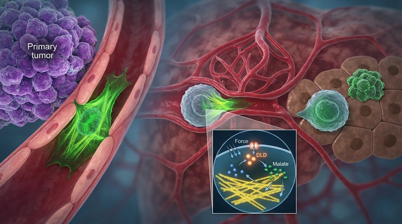
When a cancer cell is mechanically compressed while navigating a tight space, a protein called hnRNPA0 detects the compression and binds to the messenger RNA that carries instructions for making DLD. This binding stabilizes the message, preventing its degradation and causing more DLD protein to be produced specifically in cells that are experiencing confinement. Cells in open spaces showed no such increase.
Elevated DLD then does something unexpected. Rather than simply producing more energy for the compressed cell, it drives an increase in a metabolite called malate through the tricarboxylic acid cycle. Malate accumulates and then binds directly to a structural protein called TUBA1B, which is a component of microtubules, the internal scaffold that gives cells their shape and allows them to push and squeeze through tight spaces.
When malate binds to TUBA1B, it promotes the assembly of microtubules. The cell’s internal skeleton is reinforced precisely when and where the cell is being physically squeezed. It becomes mechanically stronger in response to mechanical stress, in the same way that bone becomes denser in response to the forces placed on it.
The result is a positive feedback loop. Compression triggers DLD production. DLD drives malate accumulation. Malate reinforces microtubules. Reinforced microtubules make the cell better at navigating compression. Better confined migration means the cell encounters more compression and continues producing more DLD.
“We found that compression-induced metabolic reprogramming fuels the cytoskeletal remodeling that cancer cells need to navigate physically confining spaces,” the researchers write. “This represents a previously unrecognized interface between mechanical forces and metabolic adaptation in the context of metastasis.”
When the researchers broke the chain
To confirm that each link in this molecular chain was genuinely necessary, the team disrupted it at multiple points.
They engineered cancer cells in which the binding site on DLD messenger RNA was deleted, preventing hnRNPA0 from stabilizing it under compression. In these cells, DLD did not increase during confined migration, malate did not accumulate, microtubules did not reinforce, and metastasis was significantly suppressed in animal models.
They also specifically disrupted the interaction between malate and TUBA1B. When this interaction was blocked, even cells with high DLD activity could not reinforce their microtubules during confinement, and metastatic spread was again reduced.
Both interventions left the primary tumor largely intact. What was impaired was specifically the machinery of spreading, confirming that the DLD-malate-TUBA1B pathway is specifically required for the confined migration phase of metastasis rather than for tumor growth in general.
The human evidence
The findings from cell lines and animal models were confirmed in human patient data. When the researchers examined tissue samples from colorectal cancer patients, they found that DLD expression was consistently elevated in tumor cells located within capillaries of primary tumors — precisely the cells that were actively in the process of migrating through confined spaces to reach distant organs.
More strikingly, the level of DLD expression in these capillary-embedded tumor cells correlated directly with the risk of metastatic recurrence in the patients whose tissue was analyzed. Patients whose primary tumor cells showed higher DLD expression in capillaries were more likely to subsequently develop metastatic disease.
This correlation does not prove that DLD causes the recurrence, but it places the finding in the right location, in the right cells, with the right clinical association to take seriously as a potential prognostic marker and therapeutic target.
A drug that already exists
One of the most immediately significant aspects of the discovery is that an inhibitor of DLD already exists. The compound 5-methoxyindole-2-carboxylic acid, known as MICA, is a well-characterized and reversible DLD inhibitor that has been studied in other contexts. In the current study, pharmacological treatment with MICA suppressed colorectal cancer metastasis in animal models by impairing the cancer cells’ ability to migrate through capillaries and endothelial gaps.
This does not mean MICA is ready for clinical use as an anti-metastatic treatment. Animal results frequently fail to translate to humans, the appropriate dose and safety profile in cancer patients would require extensive testing, and the drug’s effects on the many other biological processes that DLD participates in would need to be carefully evaluated. DLD is involved in normal energy metabolism throughout the body, and blocking it systemically carries potential risks that the current study was not designed to address.
What the pharmacological experiment establishes is that the pathway is druggable in principle. A target has been identified, a tool compound exists, and the biological rationale for pursuing clinical development is now in place.
What this changes about how we think about metastasis
The dominant framework for understanding metastasis has long focused on genetic mutations: cells that have acquired the right combination of mutations can survive in the bloodstream and seed distant tumors. This framework is correct as far as it goes. But it cannot fully explain why some cells within the same tumor metastasize and others do not, or why the act of traveling through the body sometimes makes cancer cells more aggressive than they were before.
The new study adds a layer that the mutation-centric view has difficulty accounting for: the mechanical environment of metastasis actively shapes the cancer cells that survive it. Being squeezed through a capillary is not simply a gauntlet that pre-adapted cells pass through. It is a training process that activates a specific metabolic program, reinforces the cells that experience it, and selects for cells that are better equipped to do it again. The physics of the body is participating in making cancer more dangerous.
This view has significant implications for how anti-metastatic therapies might be designed. Targeting the genetic changes that predispose cells to metastasize addresses one dimension of the problem. Targeting the mechanical adaptation machinery that cancer cells activate during transit addresses another. The DLD pathway represents the first clearly defined molecular example of the second kind of target, and the evidence suggesting it is relevant in human patients makes it a credible place to start.
Source
Min Liu, Bing Liu, Chen Chen, Yi-Ran Wang, Xiaoyan Li, Yajuan Zhang, Xinyang Liu, Dingpei Zhou, Hong Gao, Yijun Qi, Chen Su, Dong Gao, Yun Zhao, Yan-Jun Liu, Quanlin Li, Weiwei Yang. “Compression-induced metabolic adaptation drives confined tumor cell migration and distant metastasis via malate-dependent microtubule reinforcement.” Cell Research, 36, 513-530, 2026.
DOI: 10.1038/s41422-026-01254-4