Scientists finally have a biological explanation for why long COVID patients lose motivation and can’t think clearly. They scanned their brains and found the dopamine system was physically damaged

Since the first wave of long COVID cases emerged in 2020, patients have described a remarkably consistent set of experiences. A profound flatness where motivation used to be. Words that no longer come easily. Movements that feel slowed, as if the body is operating through resistance. A mental fog that does not lift even after months of rest. These symptoms have disrupted careers, relationships, and quality of life for millions of people. They have also proved maddeningly difficult to explain in biological terms.
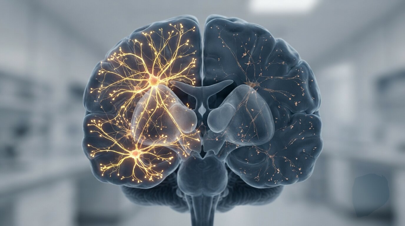
A new study published today in eBioMedicine, part of the Lancet portfolio and one of the most rigorous journals in clinical medicine, provides what researchers describe as the clearest physical evidence yet of what long COVID is doing to the brain. Using the same imaging technology used to detect early Parkinson’s disease, a team led by Jeffrey Meyer at the University of Toronto scanned the brains of 24 long COVID patients and 24 healthy matched controls. They were not looking for general inflammation or nonspecific brain changes. They were measuring one specific thing: the health of the neurons that release dopamine.
What they found was a measurable, consistent, and symptom-correlated loss of dopamine nerve terminals averaging 18 percent across the long COVID group.
What the scans actually measured
Dopamine is a chemical messenger that regulates motivation, movement, learning, and the experience of reward and pleasure. It is produced in specific clusters of neurons deep in a brain structure called the striatum, and its release depends on tiny molecular pumps called vesicular monoamine transporter 2 proteins. These pumps package dopamine into cellular sacs so it can be fired across synapses to communicate with neighboring cells.
The reason this protein matters for the study is precise: it is almost exclusively found in dopamine-releasing neurons in the striatum. Measuring how much of it is present gives researchers a highly accurate count of how many intact dopamine nerve terminals remain. The team used a radioactive tracer that binds to this protein, injected it into participants, and tracked where it accumulated using positron emission tomography.
In the 24 long COVID patients, the tracer found significantly less protein to bind to than in the healthy controls. Across three key regions of the striatum, the long COVID group showed an average loss of 18 percent of dopamine terminals. Meyer described the magnitude of this loss as clinically meaningful: comparable to the kind of damage seen in other neurological conditions where symptoms are well-established and taken seriously by clinicians.
“The magnitude of loss is about 18% of the dopamine nerve terminals on average,” Meyer told PsyPost. “In other illnesses this magnitude of loss is associated with symptoms: loss in one region is associated with trouble with motivational energy problems, loss in another region is associated with some slowness of movement, and loss in a third region is associated with memory trouble.”
The damage mapped directly onto the symptoms
The most striking finding in the study was not the average loss of dopamine terminals but the precision with which specific regional losses predicted specific symptoms.
In the ventral striatum, a region that processes motivation and the anticipation of reward, lower dopamine terminal density correlated directly with higher scores on a validated apathy scale. The same patients who showed the most damage in this area reported the greatest loss of motivational energy in their daily lives.
In the dorsal putamen, a region heavily involved in controlling physical movement, lower terminal density correlated with slower performance on a standardized finger-tapping test. Patients with more damage in this region moved measurably more slowly.
In the dorsal caudate, a region that supports learning and memory encoding, lower terminal density correlated with poorer scores on a delayed verbal memory test. Patients with the most damage in this region struggled most to retain new information.
“The correlations in loss of the marker of dopamine nerves with symptoms were stronger than expected and correlated with a wider range of symptoms than expected,” Meyer said.
This level of anatomical specificity is important. It rules out the possibility that the PET findings are a general artifact of illness or low mood. The damage is not diffuse. It is regionally organized in a pattern that mirrors the functional architecture of the dopamine system, and it tracks with the precise symptom profiles of individual patients.
How COVID may be destroying dopamine neurons
Meyer’s path to this finding began with an earlier observation. In prior long COVID brain imaging work, he found that the areas of greatest inflammation overlapped with the brain’s dopamine pathways. That overlap, combined with a separate piece of biological evidence, led him to suspect the dopamine system specifically.
The COVID-19 virus enters cells by binding to a receptor protein called ACE2. Research has established that the cells which produce dopamine carry unusually high concentrations of this receptor compared to other brain cells. This means dopamine-producing neurons may be disproportionately vulnerable to direct viral infection, and separately, that the inflammation triggered by the immune response to the virus may concentrate in regions dense with dopamine circuitry.
“Sometimes inflammation can damage dopamine releasing nerves,” Meyer said.
Which of these two pathways, direct viral damage or inflammation-mediated damage, is responsible for the 18 percent loss remains unclear. What the study establishes is the endpoint: the terminals are gone, the loss is measurable, and it correlates with the symptoms patients have been reporting for years.
The researchers also tested blood samples from participants to see whether simple biomarkers could reflect the same damage. They found no significant correlations between blood markers and the brain imaging data. This has a direct practical implication: blood tests alone are not sufficient to detect or measure this type of neurological damage in long COVID patients.
What this means for treatment
The clinical implications of the finding depend on a question the study cannot answer: whether the loss of dopamine terminals is permanent or reversible.
Meyer is explicit that it may not be permanent for everyone. The nervous system retains some capacity for repair, and some patients who recover from long COVID may regenerate terminals without intervention. But for patients whose symptoms persist without improvement, the study suggests a concrete biological target.
“People with long COVID with symptoms of low motivational energy, slowed speed taking longer to complete activities and problems with remembering words probably have lost nerves that release a chemical called dopamine,” Meyer said. “Some people may grow new nerve terminals and recover but for those who do not, there is an opportunity to make treatments to help nerves either release more dopamine or regrow nerve terminals.”
Several existing medications already approved for other conditions work by preventing dopamine breakdown or providing chemical precursors that the brain converts into dopamine. These drugs are already in clinical use for Parkinson’s disease and related conditions. Meyer’s team is now in the process of seeking approval for a clinical trial that would test whether one such medication can alleviate the motivational and cognitive symptoms of long COVID by targeting exactly the mechanism this study has identified.
“I am close to receiving approval for a clinical study to repurpose a medication for long COVID,” Meyer said. “The medication would help nerves release more dopamine and lower some types of brain inflammation. We hope it will help with memory problems and difficulty with motivational energy.”
The study has real limitations. The sample of 24 long COVID patients is small, and all participants had developed significant neuropsychiatric symptoms. Patients whose long COVID presents primarily as respiratory or cardiovascular problems may show a different or absent pattern of dopamine damage. The PET scans measure the density of transporter proteins rather than the neurons themselves, which means it is theoretically possible that the neurons are structurally intact but have reduced their protein production without actually dying.
Despite these caveats, the study represents a meaningful shift in how long COVID’s neurological effects can be understood and communicated. For years, patients with these symptoms have occupied an uncomfortable position in medicine: their suffering is real, their functional impairment is measurable, but the biological story behind it remained incomplete. A PET scan showing 18 percent fewer dopamine terminals, organized in a pattern that matches specific symptoms with anatomical precision, is not an incomplete story.
The study “Loss of vesicular monoamine transporter 2 in striatum of long COVID and relationship to neuropsychiatric symptoms” was authored by Yuhan Karida Liu, Jeffrey H. Meyer, and colleagues at the University of Toronto and the Centre for Addiction and Mental Health, and published July 30, 2026 in eBioMedicine.
Source: University of Toronto / Centre for Addiction and Mental Health. DOI: 10.1016/j.ebiom.2026.106339