Researchers scanned 47,000 brains and found that the most overlooked structure in neuroscience may be the strongest protection against cognitive decline and Alzheimer’s

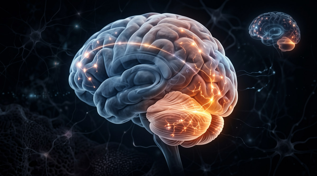
When neuroscientists discuss what determines whether a person stays mentally sharp in old age, the conversation almost always centers on the same structures: the hippocampus, where memories are formed and stored, and the prefrontal cortex, where executive control and planning live. These two regions dominate the research literature on cognitive aging, and for good reason. They are where the most visible damage accumulates in Alzheimer’s disease, and they are where volume loss correlates most strongly with the symptoms that drive people to a doctor.
What happens in the cerebellum during aging has been treated as a secondary question, if it has been treated as a question at all. The cerebellum, the dense, ridged structure at the base of the skull, contains more neurons than the rest of the brain combined and has been understood primarily as a motor coordination system, the part of the brain that makes movement smooth and automatic. Its involvement in cognition has been acknowledged for years but largely set aside in discussions of age-related decline. It was not where the action was thought to be.
A study published in Nature Neuroscience has changed that picture in a way that is difficult to dismiss. Drawing on brain imaging data from 47,144 adults across three major research cohorts, a team led by Princeton Neuroscience Institute researchers found that cerebellar volume is one of the strongest predictors of cognitive resilience in aging they measured, stronger than hippocampal volume, stronger than prefrontal cortex volume, and operating through a mechanism that neither of those structures can explain.
How the cerebellum ages, and why it is not uniform
The first major finding of the study concerns the structure of cerebellar aging itself. The cerebellum is not a single homogeneous organ. It is organized into lobules, distinct anatomical segments with different connectivity patterns and different functional roles. The anterior lobules, toward the top and front of the structure, are primarily involved in motor control. The posterior lobules, particularly the regions called crus I and crus II, are densely connected to the prefrontal cortex, the parietal cortex, and the default mode network, the same broad networks that support higher cognition, attention, and memory.
When the researchers examined how these lobules aged across their combined dataset of 47,000 participants, they found a striking gradient. The posterior association lobules, crus I and crus II, lost volume at rates of up to 6.4 percent per decade. The anterior motor lobules were significantly more preserved. The same posterior-vulnerability pattern emerged from two completely independent measurement approaches: standard volumetric MRI and a specialized tissue-composition analysis sensitive to myelin density.
The finding matters because it means the cerebellum does not simply shrink uniformly with age. The parts of it that are most deeply integrated with cognitive brain networks are also the most vulnerable to aging. And those same parts, when they remain larger and better preserved, appear to be doing something actively protective.
What a larger cerebellum actually does for cognition
The core finding from the UK Biobank data, which alone included more than 45,000 adults, is the most practically significant result in the study. Across two cognitive tests measuring processing speed and executive function, participants with larger cerebellar volume performed measurably better as they aged. By age 80, individuals with above-average cerebellar volume completed a standard processing speed task approximately 5 percent better and finished a mental flexibility test roughly 9 seconds faster than those with below-average cerebellar volume.
Nine seconds on a cognitive test sounds abstract until you consider what the difference represents. The trail-making test, which the UK Biobank administered, requires participants to connect numbered and lettered circles in alternating sequence, a task that draws on processing speed, working memory, and mental flexibility simultaneously. A 9-second advantage at age 80 reflects a meaningfully different functional state, not merely a statistical difference. It is the kind of gap that, in a clinical context, distinguishes someone who is managing independently from someone beginning to struggle.
Critically, the cerebellar volume effect was not simply piggybacking on other brain structures. The researchers directly tested whether the same protective pattern could be explained by the hippocampus, the posterior cingulate cortex, the frontal cortex, or other cortical regions known to support cognition in aging. None of them showed the same interaction: when those regions were included as covariates, the cerebellar volume effect remained statistically significant and largely intact. The cerebellum was providing reserve that the cortex was not accounting for.
The Alzheimer’s data: a threshold that changes everything
The findings from the Alzheimer’s Disease Neuroimaging Initiative data introduce a more nuanced picture with direct clinical relevance. Among participants with a clinical diagnosis of Alzheimer’s disease, the researchers separated those with low amyloid burden from those with high amyloid burden using PET imaging.
In patients with lower amyloid accumulation, the early stage of the disease before pathology has spread widely, larger cerebellar volume was strongly and significantly linked to better cognitive performance. This held even among the highest-risk genetic group: people carrying two copies of the APOE ε4 gene, which is the strongest known genetic risk factor for late-onset Alzheimer’s, showed the strongest cerebellar reserve effect in the low-amyloid group.
In patients with high amyloid burden, the protective effect essentially vanished. Cerebellar volume was no longer linked to cognitive outcomes in this group.
The researchers interpret this through what they call a threshold-reserve model. The cerebellum, on this account, acts as a buffer that helps sustain cognitive function during the early stages of Alzheimer’s pathology, before amyloid has accumulated to the point where it overwhelms whatever compensation the cerebellum can provide. Once that threshold is crossed, the reserve is spent. But below it, a larger cerebellum can apparently keep a person functioning at a higher level despite the presence of disease.
One detail from the Alzheimer’s analysis adds an unexpected dimension. While neocortical volume declined steadily and significantly across diagnostic stages from cognitively normal to mild impairment to dementia, cerebellar volume did not differ across those stages. The cerebellum is not simply following the same trajectory as the cortex, getting smaller as the disease progresses. It is maintaining its volume even as the cortex shrinks. And in the patients where it matters most, where amyloid burden is still low enough for compensation to be possible, a larger cerebellum appears to be holding the line.
Why the field missed this
The researchers address directly why the cerebellum has been underrepresented in cognitive aging research despite containing more neurons than the entire cerebral cortex. Most prior studies either treated the cerebellum as a single structure, obscuring the regional differences that proved critical in this analysis, or used sample sizes too small to detect the interactions the Princeton team identified. A 708-person cohort is not large enough to find a statistically robust interaction between age and cerebellar volume on cognitive outcomes. A 45,000-person cohort is.
The scale of the UK Biobank data was essential. The interaction effect the study identified is real but not enormous. In a smaller sample, it would have been invisible in the noise. It took nearly half a million individual brain-age-cognition data points to resolve it with the precision that changes a field’s assumptions.
The finding also reinforces a broader lesson from cognitive reserve research: the brain structures that determine who stays sharp in old age are not necessarily the ones where visible damage appears. The hippocampus shrinks in Alzheimer’s. The frontal cortex thins. These are where the symptoms live. But a structure elsewhere, one that remains relatively intact even as those regions deteriorate, may be providing the reserve that determines how long symptoms are suppressed.
What can be done about cerebellar volume
The study does not identify causes of inter-individual differences in cerebellar volume, and it does not test interventions. It is a large-scale observational study, and its findings are cross-sectional: it measured cerebellar volume and cognition at the same time rather than tracking the same people through their aging.
What it establishes is that cerebellar volume variation across adults matters for cognitive aging at a scale and with a consistency that was not previously documented. The question of what builds and maintains cerebellar volume across the lifespan is now a significantly more important one than it was before this study was published. Physical exercise, which has known effects on cerebellum-dependent motor learning, is one candidate. The cerebellum’s dependence on cerebellar-cortical circuit maintenance suggests that cognitively demanding activities might also play a role. But these are hypotheses, not conclusions.
What the 47,000-person dataset does conclude is that the cerebellum is not the passive bystander it was treated as for decades of cognitive aging research. It is an active participant, one that contributes uniquely to the brain’s capacity to maintain function as pathology accumulates, and one that the field’s existing models were built around ignoring.
Source
Federico d’Oleire Uquillas, Esra Sefik, Jakob Seidlitz, Edan Daniel Hertz, Rafael Romero-Garcia, Varun Warrier, Richard A.I. Bethlehem, Aaron F. Alexander-Bloch, Jonathan D. Cohen, Samuel S.-H. Wang, Jorge Sepulcre, Patrizia Vannini, Jesse Gomez et al. “Cerebellar aging is spatially heterogeneous and supports cognitive resilience in later life.” Nature Neuroscience, 2026; 29: 1699-1710.
DOI: 10.1038/s41593-026-02289-x