A new study found that the sleep loss in Alzheimer’s is not caused by plaques. It is caused by the brain’s own immune response to them, and researchers found a way to reverse it

Sleep disruption is one of the most devastating and least discussed symptoms of Alzheimer’s disease. While the public conversation about the condition focuses almost entirely on memory loss, the sleep changes that accompany Alzheimer’s begin years before significant cognitive decline becomes apparent, and they accelerate the disease in ways that compound every other symptom. Patients lie awake for hours during what should be restorative sleep. Their caregivers, attuned to every movement and vocalization, lose sleep alongside them. The exhaustion is total, and for decades medicine has had almost nothing to offer, because the assumption was that the sleep loss came from the same source as everything else: the amyloid plaques destroying neurons throughout the brain.
A new study from the University of Kentucky’s Sanders-Brown Center on Aging has shown that assumption was wrong. The plaques are not what is keeping Alzheimer’s patients awake. Their own brain’s immune cells are. And when researchers used a drug to temporarily silence 87 percent of those immune cells in mice with Alzheimer’s pathology, more than two hours of restorative sleep returned every night, without a single plaque being removed.
The brain’s immune system turns against sleep
Microglia are the central nervous system’s resident immune cells, derived from the yolk sac and embedded throughout the brain from early development. Their normal job is surveillance: they constantly survey brain tissue, clear cellular debris, prune unnecessary synaptic connections, and mount inflammatory responses when they detect threats. They are an essential and generally protective component of brain function.
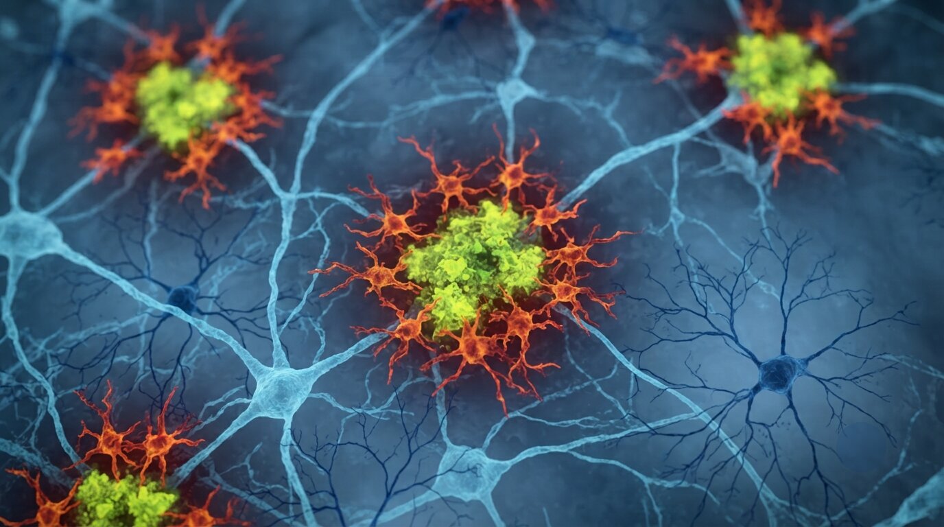
When amyloid-beta protein begins aggregating into the sticky plaques that define Alzheimer’s disease, microglia detect the accumulation and respond vigorously. They cluster around plaques, attempt to clear them, and release inflammatory signaling molecules to recruit further immune activity. The response is appropriate in principle: something foreign and potentially damaging has appeared in the brain’s environment, and the immune system is attempting to address it.
But the response that follows is disproportionate, and its collateral damage extends well beyond the plaques themselves. The inflammatory cascade that microglia initiate in response to amyloid accumulation produces a sustained state of neural excitation, a biological equivalent of an alarm system that cannot be switched off. The brain, flooded with inflammatory signals, cannot achieve the quiet neurological state that deep restorative sleep requires.
Lead researcher Shannon Macauley described the dynamic this way: “Microglia are immune cells that, when they respond to plaques, kick off this elaborate cascade of inflammation, as if the microglia are partying all night, and keeping the brain awake.”
The experiment that changed the picture
The Kentucky team studied two groups of mice: animals genetically predisposed to develop amyloid plaques, and normal aging controls. They measured both groups at six months, when plaques first begin to emerge, and at 18 months, representing late-stage disease. Each animal wore a small headmount that continuously recorded electroencephalography and electromyography, giving the researchers a precise, continuous record of when each mouse was in deep NREM sleep, REM sleep, or wakefulness. They also used light-sheet microscopy, a technique that makes brain tissue optically transparent and illuminates it with a thin laser sheet to create a three-dimensional map of every plaque and every microglial cell simultaneously.
The first unexpected result came from comparing the early and late disease time points. When the researchers looked at sleep loss and brain electrical activity in the six-month mice, where plaques had just begun to appear, and the eighteen-month mice, where plaque burden had more than doubled, they expected to find that sleep had deteriorated substantially as the disease progressed. It had not.
“I expected that as plaque burden became more severe, sleep disruption would also worsen,” said first author Nicholas Constantino. “The disruptions in sleep and cortical EEG activity that occur at six months, when plaques first emerge, did not worsen by 18 months, despite more than double the amount of plaque burden.”
The sleep deficit had reached a ceiling at the earliest stage of disease and stayed there. The damage was not being done by the plaques piling up. It was being done by the initial immune storm that the first plaques triggered, and once that storm was underway, additional plaques did not make it worse because the system was already maximally disrupted.
Two hours returned without moving the plaques
To confirm that microglia were the cause rather than a bystander, the team used a drug called PLX3397, originally developed in cancer research, which blocks the survival signal that microglia depend on. Over fourteen days of treatment, the drug depleted 87 percent of the brain’s microglial population. The inflammatory cascade collapsed. The excitatory signaling that had been driving arousal throughout the night quieted.
The mice gained more than two hours of restorative sleep per night. Their NREM sleep bouts became longer, which allowed them more time to transition into the REM sleep important for memory consolidation. Their brain electrical signatures, which had carried the distinctive fingerprint the team had identified as an early marker of Alzheimer’s, shifted back toward normal patterns.
The plaques were unchanged. Not a single amyloid aggregate had been cleared. Everything that had been building for months in the diseased brains remained exactly where it was. Only the immune response to those plaques had been interrupted, and that interruption was sufficient to restore sleep.
Why NREM sleep matters in Alzheimer’s specifically
The distinction between which sleep stage Alzheimer’s targets and which normal aging targets carries significant implications for understanding the disease. Normal aging, the researchers found, reduces REM sleep, the stage associated with dreaming and memory consolidation. Alzheimer’s pathology targets something different: NREM sleep, the deep restorative stage during which the body undergoes physical repair, the brain’s glymphatic system flushes out metabolic waste including amyloid-beta itself, and critical processes of immune regulation and cellular maintenance occur.
The consequence of losing NREM sleep in Alzheimer’s is therefore more than a quality of life issue. The glymphatic clearance that NREM sleep enables is the brain’s primary mechanism for removing the same amyloid proteins that are accumulating to form plaques. When NREM sleep is disrupted, glymphatic clearance is impaired. When clearance is impaired, amyloid accumulates faster. When amyloid accumulates faster, microglia respond more vigorously. When microglia respond more vigorously, the inflammatory cascade further disrupts NREM sleep.
“When Alzheimer’s patients lose this stage, they lose their brain’s primary cleaning cycle, creating a feed-forward loop that may drive further damage,” Macauley said.
Breaking that loop by restoring NREM sleep, even if plaques remain, could therefore have effects that extend well beyond sleep quality. Whether improved glymphatic clearance driven by restored sleep might slow plaque accumulation or reduce downstream neurodegeneration is a hypothesis the Kentucky team is now positioned to test.
The unexpected therapeutic implication
PLX3397, the drug used in this study, depletes microglia entirely, a blunt approach that would not be appropriate for long-term human use. Microglia serve essential functions throughout the brain, and their complete elimination, even temporary, would compromise immune surveillance in ways that create their own risks. The drug was a research tool, not a therapeutic candidate.
But the finding it produced has generated a more refined therapeutic hypothesis. If the problem is not the presence of microglia but their overactivated inflammatory state, then the target is not elimination but recalibration. The Kentucky team is now investigating two existing drugs with favorable safety profiles for this purpose: Metformin, the widely used diabetes medication that affects cellular energy metabolism and has shown anti-inflammatory properties in multiple contexts, and Stiripentol, an antiseizure medication with documented effects on microglial fuel use.
The goal would be to calm rather than remove the inflammatory response, reducing the nighttime excitatory signaling that prevents deep sleep without impairing the baseline immune surveillance that microglia provide. If either compound achieves this in animal models, the pathway to human trials is considerably shorter than it would be for a novel compound, since both drugs have established safety records in people.
Macauley’s team has also identified a near-term clinical application that does not require any new drug at all. The EEG signatures they documented as markers of early microglial activity in Alzheimer’s are detectable through portable, non-invasive equipment that could be used in outpatient settings and even patients’ homes.
“Portable EEG systems could allow us to monitor people in their home environments and potentially screen for changes associated with Alzheimer’s disease, without the initial need for expensive or invasive tests,” Macauley said. Local clinicians could screen at-risk patients years before cognitive symptoms appear, enabling earlier intervention with whatever tools become available.
What this changes about how Alzheimer’s is treated
The history of Alzheimer’s drug development is heavily concentrated on amyloid. The plaques were identified as the defining feature of the disease, and the dominant therapeutic strategy for decades has been to clear them: antibodies that flag amyloid for immune clearance, small molecules that prevent aggregation, drugs that reduce production. Some of these approaches have recently shown modest clinical benefits. Most have failed.
The Kentucky findings do not argue that amyloid clearance is wrong. They establish that amyloid is not the only story, and that at least one major symptom of Alzheimer’s, the sleep disruption that affects between a quarter and nearly half of all patients, is being driven not by plaques but by the immune response to them. Treating that immune response independently of plaque burden opens a therapeutic avenue that does not require first solving the amyloid problem.
For patients and families living with Alzheimer’s today, the practical significance of that avenue is immediate. Restoring restorative sleep would not reverse neurodegeneration that has already occurred. But it could reduce the daily cognitive burden of existing in a perpetually sleep-deprived state, improve the body’s capacity for self-repair during the hours it is most capable of it, and potentially slow the inflammatory cascade that the team’s ceiling effect data suggests is doing its greatest damage in the earliest stages of the disease.
The sleep loss in Alzheimer’s has been treated as a symptom of the disease’s destruction. The Kentucky study suggests it is partly a product of the brain’s own attempt to fight back, and that the fight itself has become part of the damage. Calming that response, even while the underlying pathology remains, may be one of the most accessible therapeutic levers the field has yet to pull.
Source
Nicholas J. Constantino, Shannon L. Macauley et al. “Early microglial response to amyloid plaques drives sleep loss in Alzheimer’s disease.” Alzheimer’s & Dementia, 2026.
DOI: 10.1002/alz.71579