One in four strokes has been misunderstood for decades, and a new study explains why the drugs most commonly prescribed to prevent it so often fail

Doctors have been treating one of the most common types of stroke based on a cause that new research suggests is wrong, and it may explain why the standard prevention drugs so often fail
When a patient arrives in a hospital after a stroke, one of the first questions clinicians ask is what caused it. For most strokes, the answer involves the same basic mechanism: fatty plaque has built up inside an artery, narrowing it until blood flow is blocked or a clot breaks loose and travels to the brain. This is the model that has shaped stroke medicine for decades. It is the reason aspirin is prescribed. It is the reason statins are given. It is the reason the clinical response to stroke prevention focuses so heavily on managing cholesterol and platelet activity.
For one of the most common types of stroke, new research from the University of Edinburgh suggests that model may be wrong. Not partially wrong. Fundamentally wrong in a way that explains a persistent and frustrating clinical problem: why the standard treatments for this particular stroke type so often fail to prevent a second one.
What lacunar stroke is and why it matters
Lacunar stroke accounts for roughly one in four of all strokes, making it one of the most prevalent forms of the disease. Unlike strokes caused by large clots blocking major arteries, lacunar strokes occur deep within the brain when its tiniest blood vessels, the small perforating arteries that feed the brain’s interior structures, become damaged and stop delivering blood to the tissue they supply. The resulting injury is typically small, often described as a lacune, a small cavity left by dead tissue, but its consequences are not. Lacunar stroke is a major cause of disability, a leading driver of cognitive decline, and one of the most significant contributors to vascular dementia. People who survive a lacunar stroke face elevated risk of additional strokes, including the silent strokes that occur without obvious symptoms but accumulate damage over time.
The condition that causes lacunar stroke is called cerebral small vessel disease, a term for the progressive deterioration of the brain’s microscopic blood vessel network. It is not fully understood, and despite its prevalence, it has no approved treatments specifically designed to target it.
The assumption that has shaped treatment for decades
The dominant theory about what drives lacunar stroke has long pointed toward the same mechanism responsible for most other strokes: narrowing of arteries caused by fatty deposits. If small vessels deep in the brain become blocked, the reasoning goes, it is likely because the larger arteries feeding them have narrowed through the same atherosclerotic process that blocks coronary arteries and causes heart attacks. This assumption justified treating lacunar stroke with antiplatelet drugs like aspirin, which reduce clot formation in narrowed arteries, and with statins, which reduce cholesterol and slow plaque accumulation.
Clinicians have long observed that these treatments work less well for lacunar stroke than for other stroke types. Aspirin reduces the risk of recurrent stroke in large artery disease, but its benefit in lacunar stroke has been inconsistent and modest in clinical trials. For decades this was attributed to the difficulty of treating small vessel disease rather than a question about whether the treatment was targeting the right mechanism.
The Edinburgh study suggests the latter explanation deserves more attention.
What the new research found
The research team examined 229 patients who had experienced either a lacunar stroke or a mild non-lacunar stroke. Each participant underwent detailed MRI brain imaging shortly after their stroke and again one year later. The scans allowed researchers to measure the condition of the brain’s blood vessels with precision, specifically comparing two distinct patterns of arterial change.
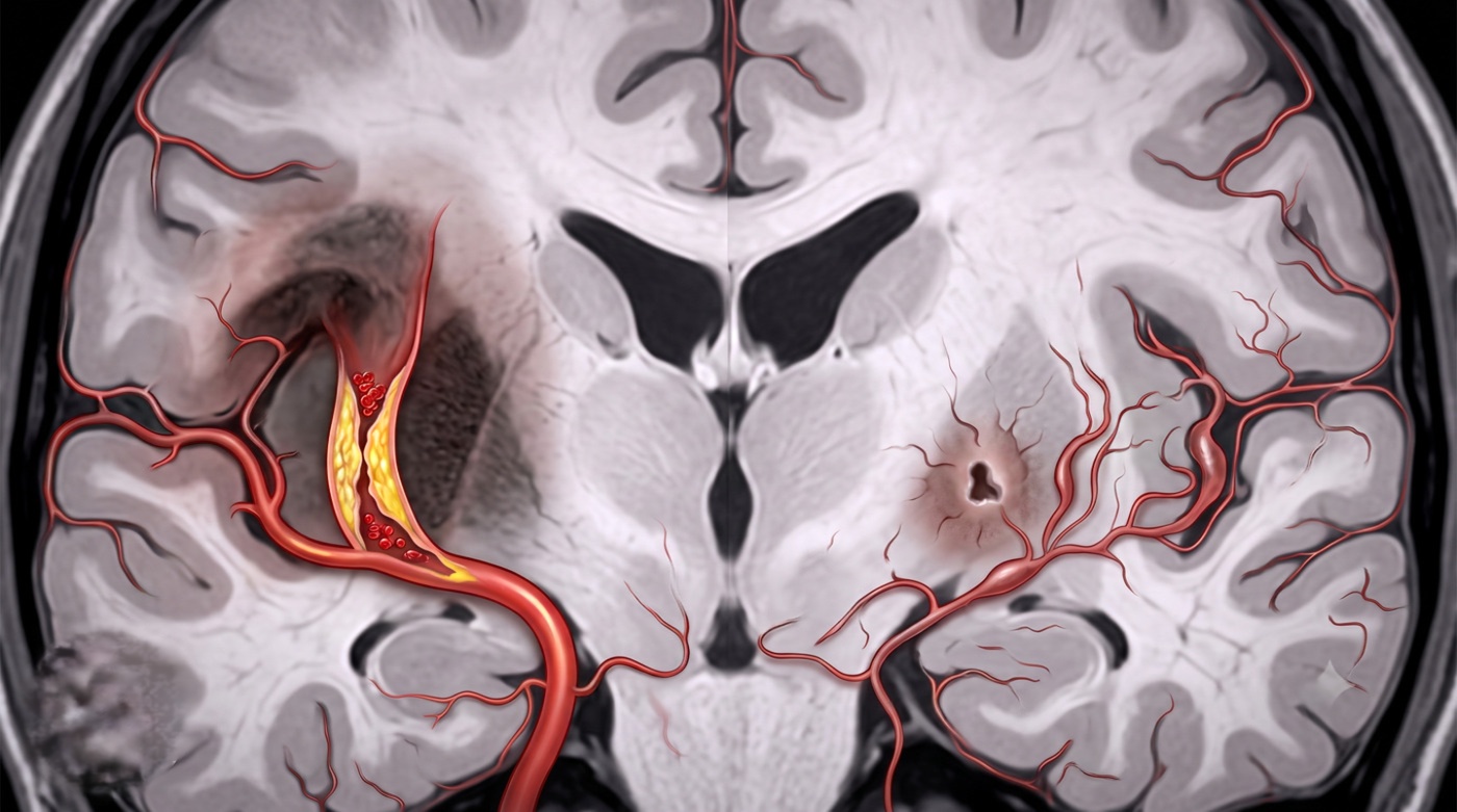
The first pattern was stenosis, the narrowing of arteries caused by fatty plaque buildup, the mechanism that underpins most standard stroke treatments. The second pattern was dolichoectasia, a condition in which arteries become enlarged, elongated, and structurally abnormal, widening rather than narrowing.
The findings separated the two stroke groups cleanly. Narrowing of large arteries was not associated with lacunar stroke. It appeared more commonly in other stroke types, exactly as existing models predicted. But artery widening showed a striking and specific link to lacunar stroke. Patients with enlarged arteries were more than four times more likely to have had a lacunar stroke than those without this pattern.
The association extended beyond the initial stroke. Artery widening was linked to more severe small vessel disease at baseline, faster progression of brain damage over the year of follow-up, and a substantially higher likelihood of developing new silent strokes during the study period. More than one in four participants developed these silent strokes during follow-up despite receiving standard stroke prevention treatments intended to stop exactly that from happening.
Why widening damages the brain differently than narrowing
The mechanism through which enlarged arteries damage small vessel function is different from the plaque-driven blockage that antiplatelet drugs are designed to interrupt. When larger arteries supplying the brain become abnormally widened and elongated, they exert abnormal mechanical forces on the small vessels that branch from them. The pulsatile pressure of blood flowing through a larger, more distensible artery is transmitted differently to downstream small vessels, potentially stressing their walls in ways that promote damage and leakage rather than blockage. This is a disease of vascular mechanics and endothelial dysfunction rather than a disease of clotting and cholesterol accumulation, and it requires a fundamentally different therapeutic approach.
“This study provides strong evidence that lacunar stroke is not caused by fatty blockage of larger arteries, but by disease of the small vessels within the brain itself,” said Professor Joanna Wardlaw, Professor of Applied Neuroimaging at the University of Edinburgh’s Institute for Neuroscience and Cardiovascular Disease and Group Leader at the UK Dementia Research Institute. “Recognising this distinction is crucial, because it explains why conventional treatments like antiplatelet drugs are not as effective for this type of stroke and highlights the urgent need to develop new therapies that target the underlying microvascular damage.”
What this means for treatment
The practical implication of the finding is not that aspirin and statins should be abandoned for all stroke patients. For strokes driven by large artery narrowing, those treatments remain evidence-based and appropriate. The implication is that for lacunar stroke specifically, the current treatment framework may be addressing the wrong target, and that developing therapies that work on the small vessel disease mechanism rather than the antiplatelet mechanism should become a clinical priority.
That work is already underway. The LACunar Intervention Trial 3, known as LACI-3, is currently evaluating whether existing medications including cilostazol, a drug that affects blood flow and vessel function, and isosorbide mononitrate, a compound used in cardiovascular disease for its effects on blood vessel dilation, can protect the brain’s small vessels, reduce recurrent stroke risk, and limit the long-term progression toward dementia and disability following lacunar stroke.
The Edinburgh study’s findings provide a mechanistic framework that supports this direction. If lacunar stroke is driven primarily by small vessel disease rooted in arterial wall dysfunction rather than plaque accumulation, then drugs that modify vessel wall function and reduce abnormal pulsatile stress should logically outperform drugs designed to prevent clotting in narrowed arteries.
The dementia connection
The study’s relevance extends beyond stroke prevention. Cerebral small vessel disease is not only the leading cause of lacunar stroke but also the underlying pathology in approximately 45 percent of all dementia cases. The white matter damage, silent strokes, and progressive tissue loss that characterize small vessel disease accumulate over years before dementia becomes clinically apparent. Understanding what drives small vessel disease more precisely, and identifying treatments that can slow its progression, is therefore not only a stroke medicine problem but one of the most important unsolved problems in dementia prevention.
The one-year follow-up data showing that brain damage continued progressing in patients receiving standard treatment underscores how inadequate the current therapeutic approach is for this population. Patients who had a lacunar stroke and received aspirin and statins continued accumulating new silent strokes at a rate that the treatments were not controlling. Whether LACI-3 and future trials targeting small vessel disease directly can change that trajectory is the central clinical question that the Edinburgh findings have sharpened considerably.
Source
Fei Han, Una Clancy, Carmen Arteaga-Reyes, Michael J. Thrippleton, Maria Del C. Valdés Hernández, Daniela Jaime Garcia, Michael S. Stringer, Ellen Backhouse, Francesca M. Chappell, Yajun Cheng, Dillys Xiaodi Liu, Junfang Zhang, Angela C.C. Jochems, Eleni Sakka, Charlotte Jardine, Gayle Barclay, Donna McIntyre, Iona Hamilton, Rosalind Brown, Yi-Cheng Zhu, Fergus N. Doubal, Joanna M. Wardlaw. “Implications of Cranial Arterial Stenosis and Dolichoectasia for Cerebral Small-Vessel Disease Etiopathogenesis: Findings From a Prospective Mild Stroke Cohort.” Circulation, 2026; 153 (23): 1813.
DOI: 10.1161/CIRCULATIONAHA.126.079493