New research found that depression is concentrated in two specific cell types in the prefrontal cortex and current drugs do not target either of them

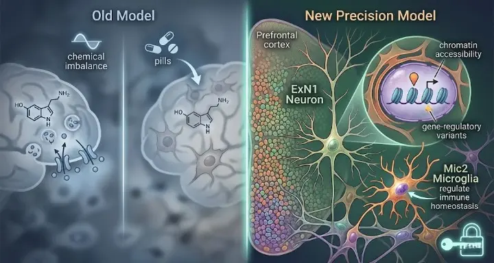
For over half a century, depression has been explained to patients using a story about neurotransmitters. Not enough serotonin. A chemical imbalance. Drugs that raise those levels help some people, which seemed to confirm the story. But serotonin-targeting antidepressants fail completely in roughly a third of patients and produce only partial relief in many more, and the neurotransmitter framework has never fully explained why. Researchers at McGill University and the Douglas Institute may have found the reason.
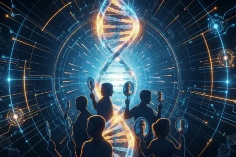
Scientists have identified two specific types of brain cells that behave differently in people with depression, offering a clearer picture of what is happening inside the brain. The finding, published in Nature Genetics, did not emerge from studying symptoms or measuring neurotransmitter levels. It came from mapping the genetic activity of more than 200,000 individual brain cells taken from the prefrontal cortex of 84 people, reading the actual molecular state of the tissue where depression lives. The result is the first precise cellular address for major depressive disorder in the history of psychiatry.
The Technology That Made This Possible
For most of neuroscience’s history, studying the brain meant studying large regions of tissue containing millions of mixed cell types, averaging together signals from neurons, immune cells, support cells, and dozens of other populations into a single blurred reading. Identifying which specific cells were behaving differently in depression from that kind of data was essentially impossible. The signal from the cells that mattered was drowned out by everything else.
The McGill team used a technology called single-nucleus ATAC sequencing, which reads the chromatin accessibility profile of individual cell nuclei at scale. Chromatin accessibility is a measure of which regions of the DNA are physically open and available for transcription at any given moment in a specific cell. When chromatin around a gene is accessible, that gene can be activated. When it is closed off, the gene goes silent regardless of what the neurotransmitter environment around the cell looks like.
By combining single-cell chromatin accessibility with gene expression in over 200,000 cells from the dorsolateral prefrontal cortex of 84 individuals, the researchers investigated gene-regulatory mechanisms underlying major depressive disorder compared to neurotypical controls. This approach does not just identify which genes are active. It maps the regulatory architecture determining which genes can be active at all, revealing a layer of biological organization that standard gene expression studies miss entirely.
The Two Cell Types at the Center
Out of every cell type in the prefrontal cortex, two showed consistent and significant differences between depressed and non-depressed individuals.
The first is a specific subtype of excitatory neuron called NR4A2-positive deep-layer excitatory neurons, which the researchers designated ExN1. These mood-related excitatory neurons showed altered gene activity in depression. NR4A2 is a transcription factor that is activated in response to stress. It regulates how neurons respond to environmental pressure, modulating the expression of genes involved in synaptic plasticity and communication. In the depressed brain, the chromatin accessibility profile of these neurons was measurably different from healthy controls, with disruptions specifically at the regulatory sites controlling genes that govern how neurons talk to each other.
The same neurons were enriched for major depressive disorder-associated genetic variants, disrupting transcription factor binding sites linked to genes that likely affect synaptic communication. This is a critical finding. The genetic variants that epidemiological studies have long associated with increased depression risk are not randomly distributed across the genome. They cluster in the regulatory regions of exactly this cell type, suggesting that genetic predisposition to depression operates specifically through the molecular machinery of these deep-layer neurons rather than through the brain’s neurotransmitter systems more broadly.
The second cell type is a specific subtype of microglia, the brain’s resident immune cells, designated Mic2. These inflammation-regulating microglia cells also show altered gene activity in depression. A gray matter microglia cluster exhibited decreased accessibility in individuals with major depressive disorder at binding sites bound by transcription factors known to regulate immune homeostasis. The immune regulatory machinery of these specific microglia was less accessible in depressed brains, meaning the cells’ capacity to maintain normal immune balance in the prefrontal cortex was compromised at the level of DNA regulation.
Why This Matters More Than It Sounds
The serotonin hypothesis of depression was never wrong in the sense of being fabricated. It was incomplete. Selective serotonin reuptake inhibitors genuinely help many people, which means serotonin dynamics are relevant to the condition. But the McGill findings point to a biological layer that serotonin-targeting drugs are not reaching.
The excitatory neurons identified in this study govern synaptic communication and plasticity in the prefrontal cortex. They respond to stress through the NR4A2 pathway. The microglia identified here regulate immune homeostasis in the same brain region. Neither of these cell populations is primarily a serotonin-dependent system. The antidepressants that target serotonin reuptake are not specifically addressing the molecular state of ExN1 neurons or Mic2 microglia. They are adjusting the broader neurochemical environment, which can have secondary effects on these cells but was never designed to target them.
The researchers found that certain neurons and microglia function differently in depressed individuals, altering brain systems tied to emotion and stress. The discovery could lead to precision therapies that target the root cellular mechanisms of depression.
The word precision is doing real work in that sentence. Current antidepressant treatment is essentially empirical: try a drug, wait six to eight weeks, assess whether it worked, try a different one if it did not. This process is driven by the absence of a precise biological target. The McGill study provides one. Treatments designed to specifically normalize chromatin accessibility in NR4A2-positive deep-layer excitatory neurons, or to restore immune regulatory function in the Mic2 microglia cluster, would be targeting depression at its actual cellular address rather than adjusting the neurochemical environment and hoping the relevant cells respond.
The Brain Bank That Made It Happen
The research depended on a resource that exists in only a handful of places in the world. The researchers used post-mortem brain tissue from the Douglas-Bell Canada Brain Bank, one of the few collections with psychiatric condition donations. Studying living brain tissue at this level of cellular resolution is not currently possible. The single-nucleus sequencing approach requires access to actual neural tissue, which means the entire field of depression neuroscience at this precision level depends on people who have chosen to donate their brains to research after death.
The 84 individuals whose prefrontal cortex tissue generated this dataset, people who lived with depression and chose in death to contribute to understanding it, produced more precise information about the cellular biology of the condition than decades of symptom-based research had managed to generate. The 200,000 cells mapped in this study represent the most granular portrait of the depressed brain that has ever been assembled.
What the Serotonin Story Got Wrong
“Depression isn’t just emotional, it reflects real, measurable changes in the brain,” said Dr. Gustavo Turecki, senior author and Canada Research Chair in Major Depressive Disorder and Suicide at McGill. “This is the first time we’ve been able to identify what specific brain cell types are affected in depression by mapping gene activity together with mechanisms that regulate the DNA code. It gives us a much clearer picture of where disruptions are happening, and which cells are involved.”
The framing of depression as a serotonin deficiency was always a simplification that served a clinical purpose, giving clinicians something concrete to target with a drug class that genuinely helped a significant proportion of patients. But it left behind the third of patients who receive no benefit from serotonin-targeting drugs, and the many more who receive only partial relief. The McGill findings suggest that for at least some of those patients, the relevant disruption is happening in excitatory neurons and microglia whose molecular state is not primarily regulated by serotonin at all.
The cellular address of depression has been found. The drugs designed to reach it do not exist yet. But for the first time, researchers know which door they need to open.
Source:
Chawla, A., Cakmakci, D., Fiori, L.M., et al. Single-nucleus chromatin accessibility profiling identifies cell types and functional variants contributing to major depression. Nature Genetics, 2025; 57(8): 1890. DOI: 10.1038/s41588-025-02249-4 https://www.nature.com/articles/s41588-025-02249-4 https://www.mcgill.ca/newsroom/channels/news/study-linking-depression-specific-altered-brain-cells-opens-door-new-treatments-366810