Depression does not just change how you feel. A study of 495,000 brain cells found it physically disrupts the brain’s ability to create new neurons in the region responsible for emotional memory.

The dominant model of depression has centered on neurotransmitters for more than fifty years. When serotonin levels drop in the synapses between neurons, mood follows. When antidepressants prevent serotonin from being reabsorbed too quickly, mood can recover. The model is supported by decades of pharmaceutical evidence: drugs that raise serotonin help many people with depression, which has seemed to confirm the underlying story.
A new study published in Nature Medicine, the most selective clinical research journal in medicine, suggests the story is correct as far as it goes and significantly incomplete beyond that.
Researchers led by Maura Dupont, a professor of psychiatry at Columbia University Irving Medical Center, assembled the largest dataset of human hippocampal tissue ever constructed for research. They collected brain tissue from people with major depressive disorder who had not been taking psychiatric medication at the time of death, and from neurotypical controls with no psychiatric diagnoses. The tissue came from three separate sources, including the New York State Psychiatric Institute brain bank and the National Institute of Mental Health Human Brain Collection Core.
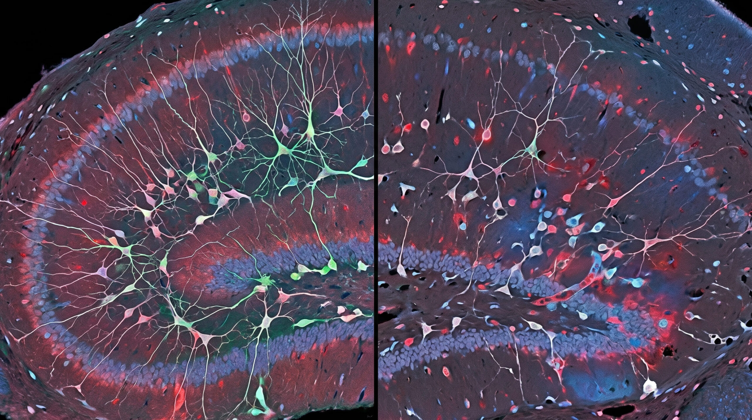
From that tissue, the team profiled 495,037 individual brain cell nuclei using single-nucleus RNA sequencing, which captures which genes are active in each cell at a given moment. They supplemented this with chromatin accessibility sequencing, which reveals which parts of the genome are structurally open and available for transcription. Spatial transcriptomics mapped where in the tissue different cell types were located and how they related to each other. Regional proteomics measured actual protein levels rather than just gene activity.
The result is described by the researchers as the largest multimodal atlas of the adult human hippocampus ever assembled. What it revealed about depression was not confined to one pathway or one molecule.
What the depressed brain was not doing
The hippocampus is one of the only regions of the adult brain that continues producing new neurons throughout life. This process, called adult neurogenesis, is thought to play a role in pattern separation, the brain’s ability to distinguish between similar memories and prevent them from bleeding into each other. When pattern separation works properly, you can remember two similar events as distinct, and carry each memory with its correct emotional weight rather than letting the emotions of one contaminate the other.
In the depressed brains, this process had stalled.
The team identified a neurogenic lineage in the adult human hippocampus: a sequence of cellular stages through which new neurons progress from stem cells to mature, integrated neurons. In the depressed brains, cells in this lineage were arrested at intermediate stages. They had committed to becoming neurons but had not completed the journey. The neurogenic process was disrupted at the transcriptional level, meaning the genes required to drive cells forward through development were not being activated in the normal sequence.
“Historically, depression was thought to be a disease of neurotransmitter deficiency, especially serotonin, but we now think that depression stems from multiple issues that affect our neurons’ ability to adapt to stress and changing environments,” Dupont said.
The stalled neurogenesis appears to be connected to a specific type of regulatory disruption. The genes that were abnormally active in the arrested cells included interferon-signaling genes and stress-response genes, suggesting that chronic inflammatory and stress signals may be disrupting the normal developmental trajectory of new neurons before they can mature.
What was happening across every other cell type
The neurogenesis finding was the most striking single discovery, but the study’s multimodal approach revealed something broader: the disruption in the depressed hippocampus was not confined to the neurogenic lineage.
Every major cell class in the hippocampus showed molecular changes in the depressed brains. Excitatory neurons, the cells that send activating signals across synaptic connections, showed dysregulation in the transcription factor networks that control their activity states. Inhibitory neurons, which normally dampen and regulate excitatory signaling, showed complementary disruptions that together suggest an altered balance between activation and suppression in the hippocampal circuit.
Astrocytes, which provide metabolic support to neurons and regulate the chemical environment around synapses, showed changes in energy metabolism and stress-response pathways. Oligodendrocytes, which produce the myelin sheaths that insulate neurons and allow signals to travel efficiently, showed disrupted gene expression. Even the cells lining the blood vessels that supply the hippocampus were altered in the depressed brains.
“The hippocampus is important for our ability to distinguish between similar but different memories and separate the emotional connotation of past memories and current events,” Dupont said. “When this ability is impaired, memories together with their emotional value become less distinct and more likely to blend together.”
The practical consequence is something that people with depression frequently describe without knowing the neuroscience behind it: a sense that past experiences of failure, rejection, or loss contaminate the present. Each new difficulty feels like confirmation of every previous one, not because the person is being irrational but because the circuit that should be keeping those memories separate is not functioning normally.
Why medication history matters for understanding the findings
The study’s decision to focus specifically on people who were not taking psychiatric medication at the time of death is methodologically important and worth explaining.
Antidepressants, mood stabilizers, and antipsychotics all affect gene expression in the brain. If the tissue samples had come from medicated individuals, it would be impossible to know whether the molecular changes observed reflected the disease itself or the effects of treatment. By restricting the study to unmedicated individuals with a confirmed clinical diagnosis of major depressive disorder, the team could be more confident that what they were seeing represented the biology of the disease in its natural state.
This restriction also creates a limitation: the people in the study represent a specific subset of individuals with depression, those whose disease was severe enough to receive a formal diagnosis but who had not been treated with medication. Whether the same molecular changes appear in people who do have access to and use antidepressant treatment, and whether treatment reverses or modifies these changes, are questions this study cannot answer.
Prior research has suggested that some antidepressants, particularly those in the SSRI class, may promote neurogenesis in the hippocampus as part of their therapeutic mechanism. If that is correct, the stalled neurogenesis the study observed in unmedicated individuals could be one of the biological pathways through which antidepressants work, independent of their more immediate effects on serotonin signaling. This would represent a significant expansion of the mechanistic understanding of antidepressant action rather than a contradiction of it.
What the findings mean for how depression is understood
The serotonin hypothesis of depression emerged in the 1960s and became dominant partly because it was testable and partly because drugs targeting serotonin worked. Both of those facts remain true. SSRIs help a substantial proportion of people with depression, and serotonin signaling is genuinely disrupted in the disease.
What this study adds is evidence that serotonin is one thread in a much more complex biological picture. The disruption in the depressed hippocampus extends to neurogenesis, to excitatory-inhibitory balance, to cellular energy metabolism, to the integrity of the myelin around neurons, and to the inflammatory state of the tissue itself. These are not secondary effects of serotonin dysregulation. They appear to be independent molecular signatures of a disease that acts on multiple systems simultaneously.
“Our discovery of a human hippocampal neurogenic lineage and evidence of stalled neurogenesis in MDD challenges the conventional neurotransmitter-focused view of this disorder,” the researchers wrote. “It positions hippocampal neurogenesis as a key therapeutic target and provides a framework for developing treatments that extend beyond serotonin pathways.”
The practical significance for treatment is still speculative but directionally important. If impaired neurogenesis is a core feature of depression rather than a side effect of other changes, then therapies that specifically promote new neuron formation in the hippocampus could represent a new therapeutic direction. Several interventions already known to promote hippocampal neurogenesis in animals and humans, including physical exercise, certain types of ketamine, and electroconvulsive therapy, are also effective treatments for depression in some patients. The neurogenesis finding provides a possible mechanistic explanation for why.
What the study cannot establish
The study examined postmortem brain tissue, which means it captures a snapshot of the brain at one moment in time rather than tracking changes as depression develops, progresses, or remits. It is impossible to tell from this data whether the stalled neurogenesis preceded the depression, appeared as the disease developed, or represented a chronic state that had persisted for years.
The samples, while the largest ever assembled for this type of analysis, came from a relatively small number of individuals: 11 with major depressive disorder and 19 controls. The sample sizes necessary for single-nucleus sequencing at this resolution are constrained by the difficulty of obtaining post-mortem human brain tissue with sufficient quality, but the small n means that individual variation between donors may have influenced the results.
The team also could not measure how long each person had been depressed, how severe their depression was at various points in their life, or whether they had experienced periods of remission. These clinical variables could meaningfully affect the molecular state of the tissue.
What the study provides, at an unprecedented level of cellular and molecular resolution, is a map of what the depressed human hippocampus looks like. The map shows more disruption, across more systems, than the serotonin model of depression would predict. Whether treating that disruption directly can help people with depression is the question that follows from this work, and it is one the field is now positioned to begin answering.
The study, “Dysregulated adult hippocampal neurogenesis in major depressive disorders”, was authored by Madeleine S. Peng, Jialin Jiang, Lucia Polizzi, and colleagues at Columbia University Irving Medical Center and New York State Psychiatric Institute, and published August 21, 2026 in Nature Medicine.
Source: Columbia University Irving Medical Center. DOI: 10.1038/s41591-026-04571-8