Scientists have known nighttime light raises cardiovascular risk. A study of 11,071 people just showed for the first time what it is actually doing to the structure of the heart.

The relationship between light at night and cardiovascular disease has been building in the scientific literature for years. Population studies have found that people living in areas with more light pollution face higher rates of coronary artery disease, heart attack, and stroke. Studies using wrist-worn light sensors in the UK Biobank found associations between brighter bedrooms and higher rates of heart failure and atrial fibrillation. The biological mechanisms proposed have centred on circadian disruption: light at night suppresses melatonin, shifts the timing of cortisol and blood pressure cycles, and keeps sympathetic nervous system activity elevated during hours when the body is designed to down-regulate.
None of those studies looked directly at what nighttime light was doing to the physical structure of the heart. They measured disease outcomes, which appear at the end of a long pathological process. The structural changes that precede those outcomes, the ones that would reveal the mechanism and the timeline, had never been mapped in a large human population.
A study published September 10 in the European Heart Journal, the flagship journal of the European Society of Cardiology and one of the most selective cardiovascular journals in the world, has done that mapping.
Researchers led by Professor Lu Qi of Tulane University recruited 11,071 participants from the UK Biobank and equipped each one with a wrist-worn light sensor for seven days. The sensors measured actual personal light exposure during sleep hours, providing a more precise measure of individual bedroom light levels than the satellite-derived estimates of light pollution used in most prior studies. Three years after the light measurements were taken, each participant returned for a cardiac magnetic resonance imaging scan, the gold standard tool for measuring the structure and function of all four chambers of the heart.
The question the study asked was direct: do people who are regularly exposed to more light during sleep have differently structured hearts?
The answer was yes, and the differences were consistent across every cardiac chamber examined.
What the cardiac MRI revealed
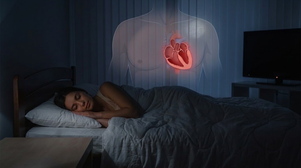
The study compared people exposed to nighttime light levels above 3 lux against people exposed to essentially no light during sleep. Three lux is a low threshold. It is roughly the level produced by a dim nightlight, the standby LED on a television, the glow from a streetlight through thin curtains, or a phone placed face-up on a bedside table.
In people regularly exposed to light above this threshold, the wall of the left ventricle was measurably thicker. The left ventricle is the heart’s main pumping chamber, responsible for pushing oxygenated blood out to the body with each beat. Thickening of its walls reduces the volume inside the chamber, meaning the heart pumps less blood with each contraction.
The heart muscle’s ability to flex normally during each heartbeat was also impaired. Myocardial strain, the technical measure of how much the heart muscle deforms and recovers with each beat, was reduced in multiple directions: circumferential, radial, and longitudinal. Each of these represents a different axis along which the heart muscle moves when it contracts and relaxes. Reduced strain in all three directions indicates that the muscle is less compliant and less efficient than it should be.
Changes were also detected in the right ventricles and left atria, the other two cardiac chambers. The pattern was consistent and dose-dependent: higher nighttime light exposure was associated with more pronounced cardiac changes. There was no threshold below which more light was clearly safe. The relationship was graded.
“This is the first study of its kind, and it shows that exposure to higher levels of light at nighttime is linked to cardiac remodelling,” said Professor Qi. “This is where the structure and function of the heart changes, and we typically see it in response to chronic stress or injury to the heart. It’s our body’s way of adapting to that stress, but ultimately it weakens the heart and can lead to heart failure.”
What cardiac remodelling means
The term remodelling describes a specific biological process that the heart undergoes in response to sustained stress. When the heart faces increased workload, whether from high blood pressure, chronic infection, metabolic disease, or any other persistent demand, it adapts by changing its physical structure. The walls thicken. The chambers enlarge or shrink depending on the type of stress. The muscle composition changes. The electrical properties of the tissue shift.
In the short term, remodelling is adaptive. A heart that thickens its walls can maintain blood pressure against increased resistance. But remodelling that persists becomes maladaptive. A thicker wall is stiffer. A stiffer wall fills less efficiently between beats. The volume ejected with each contraction decreases. Over years, this process leads to diastolic dysfunction, the inability of the heart to relax and fill properly, and eventually to heart failure.
The structural changes documented in the 11,071 participants with higher nighttime light exposure are the early markers of this process. They are not disease outcomes yet. They are the biological precursors that, if they progress, produce the disease outcomes that prior population studies have already documented in light-exposed populations.
The study also examined outcomes in a parallel analysis of more than 73,000 UK Biobank participants followed for 8 to 10 years. In this larger sample, higher nighttime light exposure was associated with 29% higher risk of heart failure and 24% higher risk of coronary artery disease, with the risk gradient following the same dose-response pattern seen in the structural imaging data.
The combination of structural data from cardiac MRI and outcome data from long-term follow-up creates an unusually complete picture. It connects the light in the bedroom to the change in the heart wall to the disease endpoint years later, tracing the chain from exposure to mechanism to outcome in a way no prior study had achieved.
The mechanism: how light disrupts the heart
The biological pathways through which nighttime light produces cardiac remodelling are not yet fully established, but the leading candidates are well-grounded in prior research.
The circadian system, the body’s internal 24-hour clock, regulates the timing of virtually every physiological process, including the cardiovascular system. Blood pressure follows a circadian rhythm, naturally falling during sleep in a pattern called nocturnal dipping. Heart rate slows. The autonomic nervous system shifts toward parasympathetic dominance, the rest-and-repair mode. Inflammatory markers cycle to their daily minimum. Growth hormone is secreted.
Light at night disrupts all of these processes simultaneously. It suppresses melatonin, the hormone that signals darkness to the rest of the body. It delays the circadian phase, pushing the timing of physiological processes out of alignment with the sleep-wake cycle. It maintains sympathetic nervous system activation at levels appropriate for wakefulness during hours when the body is designed to recover.
Chronically elevated sympathetic activity during sleep is one of the most direct routes to cardiac remodelling. The sympathetic nervous system drives the release of norepinephrine, which has direct effects on heart muscle cells. At sustained high levels, norepinephrine promotes cardiac fibrosis, thickens ventricular walls, and impairs the electrical properties of heart tissue. These are precisely the changes the MRI data documented.
The dose-response relationship observed in the data suggests that the mechanism is cumulative. More light, more disruption, more structural change. The absence of a safe threshold below which light exposure was clearly benign is consistent with a process that operates continuously rather than switching on at a specific level of exposure.
How much light is too much
Three lux is the threshold the study used, but the researchers found dose-dependent effects rather than a binary safe-versus-unsafe cutoff. Three lux is a very low level of light. For comparison, bright indoor lighting is typically 300 to 500 lux. Twilight is roughly 10 lux. A full moon on a clear night produces about 0.1 lux. A television on standby at the far end of a room might produce between 1 and 5 lux.
For most people sleeping in contemporary bedrooms, achieving light levels below 3 lux requires specific effort: blackout curtains to block street lighting and morning sunrise, covering the standby indicators on electronics, not using phones as bedside clocks, and avoiding screens in the hour before sleep.
An editorial accompanying the paper, written by Professor Thomas Münzel of the University Medical Center Mainz and colleagues, translated the findings into direct clinical guidance. “It is time for clinicians, particularly those managing patients with heart failure or atrial fibrillation, to start asking about the sleep environment: how dark the bedroom is, whether the patient works night shifts, and how much screen use occurs after sunset. The advice is simple and inexpensive: blackout curtains, warm-coloured bedside lighting, and covering the small standby LEDs on household electronics.”
The editorial went further: “Darkness deserves recognition as a vital sign, as essential to cardiovascular health as blood pressure control and clean air.”
What the study cannot establish
The study is observational. It demonstrates that higher nighttime light exposure is associated with cardiac remodelling and later cardiovascular disease events, but it cannot prove that the light caused these outcomes. People who sleep with more light may differ from people who sleep in complete darkness in other ways that affect cardiovascular risk. Higher light exposure may correlate with living in more urbanized, noisier, more economically stressed environments that carry their own cardiovascular burden.
The seven-day light measurement provides a reliable estimate of typical sleep-time light exposure, but it does not capture variation across seasons, years, or life changes. People whose bedroom light levels shifted substantially after the measurement would not be correctly categorized by their baseline sensor data.
The cardiac MRI was also collected at a single time point, three years after the light measurement. This allows the researchers to connect prior light exposure to later cardiac structure, but it cannot track the progression of structural changes in the same individuals over time.
What the study provides, at 11,071 participants with direct objective light measurement and gold-standard cardiac imaging, is the first systematic map of what nighttime light exposure is doing to the structure of the human heart. The map shows changes that are consistent with the known biology of circadian disruption, that follow a dose-response relationship across all four cardiac chambers, and that align with the disease outcomes documented in longer follow-up studies. The practical implication is clear enough to act on without waiting for additional evidence: a darker bedroom is a healthier environment for the heart.
The study, “Nighttime light exposure and cardiac structure and function”, was authored by Jin Dai and colleagues at Tulane University, and published September 10, 2026 in the European Heart Journal.
Source: European Society of Cardiology. DOI: 10.1093/eurheartj/ehag563