New research found that people whose daily activity peaks after 2pm have a 45% higher dementia risk and the brain scans explain why timing matters more than duration

Most people think about their circadian rhythm in terms of sleep. Go to bed at a reasonable hour, wake up at a consistent time, avoid screens late at night, and the body clock takes care of itself. Two studies published in early 2026, one from UT Southwestern Medical Center and one from Johns Hopkins Bloomberg School of Public Health, suggest that this framing is missing most of the picture. The strength and consistency of your daily rest-activity rhythm, measured not just at night but across the entire 24-hour cycle, predicts whether your brain will shrink faster or slower as you age. And the habits shaping that rhythm are operating right now, decades before any neurological symptom would appear.
The findings reframe circadian rhythm from a sleep hygiene issue into something considerably more consequential. The body clock is not just regulating when you feel tired. It is regulating the biological processes that determine whether your brain retains its volume, its connectivity, and its resistance to neurodegeneration over the decades ahead.
The Numbers From 2,000 Older Adults
The UT Southwestern study, published in the journal Neurology in December 2025, followed 2,183 adults with an average age of 79 who were free from dementia at the start of the research. Each participant wore a small chest-based monitor continuously for approximately 12 days, generating objective accelerometer data about their daily patterns of rest and activity rather than relying on self-reported sleep logs. Over a median follow-up of three years, 176 participants received a dementia diagnosis.
Participants in the weakest rhythm group had nearly 2.5 times the risk of developing dementia compared with those whose rhythms were strongest, even after adjusting for age, cardiovascular risk factors, education, and genetic risk such as APOE ε4 status. Each standard-deviation decrease in rhythm strength translated into a 54% higher dementia risk.
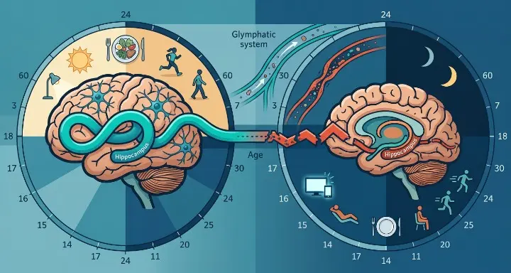
The timing of activity across the day mattered independently of rhythm strength. Individuals whose daily activity peaked later in the afternoon, after about 2:15 p.m., had a 45% higher risk of dementia compared with those whose activity peaked earlier. This finding has direct implications for people whose schedules push their most active period into the afternoon or evening, whether through work patterns, lifestyle habits, or the progressive circadian drift that comes with age.
The Brain Shrinkage Data
The Johns Hopkins study, published in April 2026, zoomed in on what is actually happening inside the brain when circadian rhythms fragment. Researchers from the Bloomberg School of Public Health examined 344 cognitively unimpaired adults drawn from the Baltimore Longitudinal Study of Aging, one of the longest-running studies of human aging in the world, with MRI data tracking changes in brain volume over time alongside accelerometer data capturing rest-activity rhythms.
Older adults with a more fragmented circadian rest-activity rhythm experienced more shrinking of the brain in areas typically affected by Alzheimer’s disease. The regions losing volume fastest in participants with weaker, more fragmented rhythms included the hippocampus and surrounding structures, the same areas that the FTL1 research, the speech timing data, and the Alzheimer’s blood test findings all converge on as the earliest sites of neurodegeneration.
The ventricular finding added a second layer of evidence. Scientists observed a correlation between a more fragmented rest-activity rhythm with a quicker increase in the volume of brain ventricles. Brain ventricles are fluid-filled spaces that often expand as surrounding brain tissue is lost. Thus, the finding suggests that more fragmented daily rhythms are associated with faster increases in ventricular size over time, which suggests a link between disrupted daily rhythms and more widespread brain atrophy.
Crucially, the Johns Hopkins team found that this relationship was not explained by sleep duration or quality alone. The circadian rhythm metric they were measuring captures the consistency and amplitude of the entire 24-hour behavioral cycle, meaning it reflects how reliably a person is active during the day and at rest during the night across days and weeks. People with high rhythm strength have clear, consistent patterns. People with low rhythm strength have irregular, fragmented cycles where activity and rest are distributed unpredictably across the day and night.
“These findings add to evidence that a weaker, more fragmented rest-activity rhythm can be an early sign of adverse neurological changes, and might also contribute to the underlying neurodegenerative process,” said senior author Adam Spira, professor in the Bloomberg School’s Department of Mental Health.
What Is Actually Regulating Circadian Rhythm Strength
The biological mechanism connecting circadian rhythm to brain health runs through multiple pathways that operate simultaneously. The glymphatic system, the brain’s waste clearance network that the abdominal pump article described, is heavily circadian-dependent. Its activity peaks during specific phases of the sleep-wake cycle and requires reliable circadian timing to function at full efficiency. When the cycle fragments, the clearance windows shorten and become less predictable, allowing amyloid-beta and tau proteins to accumulate between clearance events.
A WashU Medicine study published in Nature Aging in late 2025 identified a specific circadian clock protein called REV-ERBα that regulates tau levels in the brain directly. When REV-ERBα activity was inhibited, tau levels fell and brain tissue damage was reduced in mouse models of Alzheimer’s disease. The protein regulates NAD+, a molecule essential for energy metabolism and DNA repair. Circadian disruption suppresses NAD+ through this pathway, creating an energy deficit in neurons that compounds the structural damage visible on MRI.
At the cellular level in the hippocampus, aging disrupts the circadian regulation of proteins involved in energy metabolism, neurotransmission, and synaptic plasticity. The daily rhythms that keep these processes synchronized, producing the right proteins at the right times, weaken as circadian strength declines. The result is a hippocampus that is running on an increasingly disrupted schedule, producing proteins at the wrong times and in the wrong quantities to support the memory consolidation and cellular maintenance that healthy aging requires.
What Shapes Rhythm Strength in Practice
The research points to several behavioral and environmental factors that directly influence circadian rhythm strength in ways that are measurable and, importantly, modifiable.
Light exposure is the primary zeitgeber, the external time-setter that anchors the biological clock to the external environment. Bright light in the morning directly activates the suprachiasmatic nucleus, the brain’s master clock, resetting the circadian system to align with the solar day. Consistent morning light exposure strengthens rhythm amplitude. Irregular light exposure, particularly bright artificial light in the evening and insufficient light in the morning, is one of the primary mechanisms through which modern indoor lifestyles fragment circadian rhythms.
Physical activity timing matters independently of light. The UT Southwestern data showing that activity peaking before 2:15 p.m. correlates with lower dementia risk suggests that when you are most active during the day is itself a circadian signal. Regular, consistent activity patterns that reinforce the natural daytime-active, nighttime-rest cycle strengthen rhythm amplitude. Irregular activity patterns, long sedentary periods during the day followed by bursts of activity in the evening, weaken it.
Meal timing operates through peripheral circadian clocks in the liver, gut, and metabolic organs that are synchronized with the central brain clock through regular feeding patterns. Irregular eating, particularly late-night eating that conflicts with the body’s nocturnal metabolic downregulation, sends contradictory timing signals to peripheral clocks, producing a fragmentation of the circadian system that extends beyond sleep.
The Window That Matters Most
One of the more important details in the Johns Hopkins study is that the researchers examined these effects specifically in middle-aged and older cognitively unimpaired adults. The brain volume changes associated with weaker circadian rhythms were detectable years before any cognitive symptom would appear on a standard neurological examination.
The longitudinal findings suggest that disrupted rhythms may precede change in brain structure, raising the possibility that more fragmented or less consistent rest-activity rhythms contribute to neurodegeneration, rather than simply reflecting it. The direction of causality matters enormously for prevention. If circadian fragmentation is a consequence of early neurodegeneration, strengthening daily rhythms would be useful as a marker but not as an intervention. If circadian fragmentation is contributing to the neurodegeneration, which the longitudinal design of the Johns Hopkins study was specifically constructed to test, then the daily habits that shape rhythm strength are genuinely modifiable risk factors for brain aging.
The UT Southwestern lead researcher, Wendy Wang, said that future studies should examine the potential role of circadian rhythm interventions, such as light therapy or lifestyle changes, to determine if they may help lower a person’s risk of dementia. That trial has not been completed yet. But the structural brain data from Johns Hopkins and the dementia incidence data from UT Southwestern are pointing in the same direction: the consistency of your daily behavioral pattern is physically restructuring your brain, slowly, over years, in ways that will eventually become visible on a scan.
Sources:
1. UT Southwestern / Neurology (December 2025) Wang, W., et al. Circadian Rest-Activity Rhythm and Risk of Incident Dementia. Neurology, December 29, 2025. DOI: 10.1212/WNL.0000000000213387 https://www.insideprecisionmedicine.com/topics/precision-medicine/circadian-rhythm-disruption-linked-to-higher-dementia-risk/
2. Johns Hopkins Bloomberg School of Public Health (April 2026) Kaizi-Lutu, M., Callow, D., Spira, A., et al. Fragmented Circadian Rest-Activity Rhythms Linked to Faster Brain Shrinkage Over Time. Published April 2026. https://publichealth.jhu.edu/2026/in-older-adults-fragmented-circadian-rest-activity-rhythms-linked-to-faster-brain-shrinkage-over-time
https://www.medicalnewstoday.com/articles/brain-health-staying-more-active-during-the-day-helps-retain-brain-volume
DOI https://doi.org/10.1002/alz.71190Digital Object Identifier (DOI)
3. WashU Medicine / Nature Aging (November 2025) Lee, J., Musiek, E., et al. Inhibiting REV-ERBα reduces tau accumulation and neurodegeneration in Alzheimer’s models. Nature Aging, 2025. https://www.sciencedaily.com/releases/2025/11/251101000713.htm